Everything You Need To Know About C-Arm X-Ray Machines
Also, try tapping into DEXA scan and find out what it can do.
The fluoroscopic system has two main configurations that can be installed permanently. The first class uses the radiolucent patient examination table with a mounted under-table tube while the imaging system is installed over the table itself. The commonly referred C-arm system is used where more flexibility is required during the examination process. The best information about what is a dexa scan is available when you click the link.
How much will you be paying for a DEXA scan?
The cost for a DEXA scan depends on the location and the kind of facility that will be doing the test.
With DEXA scan, you can see how your bones are doing and if they are just right for your age because there are a bunch of young adults who are supposed to have strong and healthy bones, but because of their lifestyle, they weakened their bones faster than other adults. DEXA scan is the kind of innovation that will save lives. Be excited to our most important info about ge c arm.
If your doctor deems it medically necessary, you can have your insurance company cover the expenses for the scan, and that is how you can save some money from the DEXA scan. It is not a cheap process, but it is going to help you be aware of what you are putting your body into and get the chance to change before it is too late.
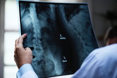
An x-ray image intensifier converts the rays into visible light that has higher intensity, more than what a standard fluorescent screen can provide. People use these intensifiers on their x-ray imaging systems to convert low-intensity light to a bright visible light that will help detect abnormalities inside a human being's body structure. Learn more details about pain management at https://en.wikipedia.org/wiki/Pain_management.
Thanks to the image intensifier, the viewer can easily see the structure of the image that is being focused by the x-ray. You should know that the XRII requires less absorbed doses since it has a more efficient conversion of x-ray to visible light.
